Dental images and CT scans for children
Most children need a panoramic image for UAH 390 rather than a CT scan. We will explain which of the two is enough in each case, so that you do not overpay and your child is not exposed needlessly.
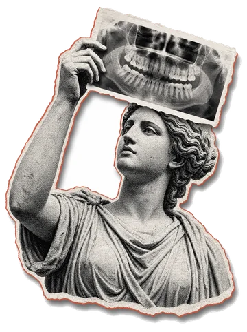 Imaging in children is first of all a question of whether it is really needed. Our position is simple: we take an image when the doctor's decision depends on it, and we take the smallest sufficient volume. Most often that is a panoramic image, not a CT scan.
When an image is genuinely needed
- Before orthodontic treatment. The most common reason. The orthodontist needs to see how many buds of permanent teeth there are, what position they are in and whether there is enough room. Without that, a treatment plan is guesswork.
- A tooth is not coming through, although the neighbouring ones have already appeared. We need to find out whether it is there at all and where it sits.
- An injury — the child has fallen and a tooth is loose or has changed colour.
- Suspected inflammation near the root of a baby tooth: it can damage the bud of the permanent tooth lying directly beneath it.
- Before the extraction of a baby tooth whose roots sit in an unusual position.
Panoramic image or CT scan
| Situation | What to do | Price |
|---|---|---|
| Before braces or a plate | Panoramic scan | UAH 390 |
| Monitoring the change of teeth | Panoramic scan | UAH 390 |
| An impacted tooth that is not erupting | CT of a segment | UAH 790 |
| A complex jaw injury | CT of a segment or of a jaw | from UAH 790 |
| Planning a complex orthodontic case | CT of both jaws | UAH 1 290 |
If the orthodontist has written “OPG”, that means a panoramic image for UAH 390 and there is no need for a CT scan. If they wrote “CBCT” or “3D”, tomography is required. If in doubt, call 067 478 87 78 and read out what has been prescribed.
The dose, honestly
The unit selects the dose itself, to match the height and build of the patient, so the figure varies: 10–15 µSv per study. The annual dose a person receives from background radiation alone is about 4000 µSv. So a CT of both jaws is roughly 0.4% of what you will receive over a year anyway, and less than a few hours on a plane.
For children the Planmeca switches on a separate mode: it reduces the scanning area to the size of a child's head and lowers the power. A child receives less than an adult does on the same protocol.
This does not mean that images can be taken just in case. It means that when the doctor has ordered one, it is not worth refusing out of fear of radiation: a problem with the bite or with a tooth bud that is not found in time costs more.
How it all happens
The child takes off everything metal and puts on an apron. One of the parents stays beside them, also in an apron. The child stands (or sits, if they are small), bites on a plastic positioner, and the unit makes one turn around the head — 20 seconds for a panoramic image. Nothing touches the face, nothing hurts, and the sound is like a quiet hum.
The one thing needed is to stay still for those 20 seconds. That is why we first show and explain everything, and only then take the image. If the child gets frightened and moves, we will redo it at no extra charge.
What you receive
A printed image to take with you and a digital copy by email — to your address and to the orthodontist's. The link never expires: when, a year later, the orthodontist wants to compare how the picture has changed, the old image will be to hand.
We take the image and hand it over — the report and the treatment plan are made by your own doctor, because the decision depends on the clinical picture they see in the chair. If you do not have a doctor of your own, you can show the image to the head doctor at Houston — with a CT of both jaws the consultation here is free of charge.
At the Houston clinic children are seen by a separate doctor — about children's dentistry.
Opening hours
Mon–Sat 8:00–19:00, Sunday 10:00–18:00. We work on Sundays, which helps when your child has school. 3B Mykhaila Hrushevskoho St., Dnipro.
On medical grounds — as a rule from the age of 5–7. It is at this age that the orthodontist wants to see the buds of the permanent teeth. Earlier than that, images are taken more rarely and only when there is no way round it: an injury, suspected inflammation, or a developmental anomaly.
The panoramic protocol for a child is about 10 µSv. The annual background dose for a person is around 4000 µSv, so the image gives a fraction of a percent of the annual background. The Planmeca has a separate children's mode and automatically reduces both the scanning area and the dose. The risk from a problem that is not found in time is higher than the risk from the image.
In most cases a panoramic image (UAH 390). It shows the buds of the permanent teeth, the order of eruption and the wisdom teeth. A CBCT is ordered more narrowly: an impacted tooth that is not erupting, a complex injury, or preparation for surgery.
Yes. One of the parents stays in the room, in a protective apron. Children feel calmer that way, and a calm child does not move — so the image comes out right the first time.
We show the child the unit, let them hear how it hums, and explain that nothing touches them and nothing hurts. The scan lasts 20 seconds. If the child is not ready, we do not push — we reschedule. A blurred image would have to be retaken anyway.
Leave a request — we will call you back during working hours and find a convenient time